Nipah virus infection gets its name from the village in Malaysia where the person from whom the virus was first isolated succumbed to the disease. Nipah Virus is an emerging infectious disease that broke out in Malaysia and Singapore in 1998 and 1999. It first appeared in domestic pigs and has been found among several species of domestic animals including dogs, cats, goats, horses and sheep. The infection is also known to affect human beings. The organism which causes Nipah Virus encephalitis is an RNA or Ribonucleic acid virus of the family Paramyxoviridae, genus Henipavirus, and is closely related to Hendra virus.
Throughout the history of the world humans have been plagued by diseases of various types and origins. Zoonotic diseases, or diseases which have the capability to jump species, animals to humans or vice versa, have been particularly troublesome and deadly. Zoonotic diseases are unique in that they are mainly caused by pathogens such as fungi, bacteria, parasites, or viruses. These pathogens typically survive in a reservoir host, which have immunity to the pathogen. The list of possible reservoir hosts capable of transmitting disease to humans is expansive; however the most common are apes, insects, rodents, and bats. The diseases are then passed to humans who come in contact with an infected animal through bites or scratches, an infected animal’s environment, or animal secretions such as saliva, feces, or mucus. Often these diseases have a higher virulence because of the lack of any immunity within the human population and the ease of transmission. Some more infamous zoonotic diseases are West Nile, Rabies, Ebola, and Dengue fever. As of recent, more and more zoonotic diseases are emerging because of an increase in human and wildlife interaction. An increase in farming and or deforestation has resulted in humans and wildlife into the same habitat. A prime example of this is the emergence of the Nipah virus (NiV). NiV is a member of the Henipavirus genus in the Paramyxoviridae, and has become a growing concern of Southeast Asia and Australia. Nipah virus is also a growing concern for the United States. The Center of Disease Control (CDC) has declared it a biosafety level 4 agent 6. This the highest biosafety level category, home to agents which can be distributed via aerosol transmission and have no treatment or vaccine. Similar biosafety level 4 agents are Ebola, Smallpox, and several hemorrhagic diseases. The CDC has also tagged the Nipah virus a Category C bioagent, the third highest priority agent category in regards to biological warfare 6. The availability, simplicity to produce and disperse, and high mortality rate of the Nipah virus make it possible for it to be used as a weapon of biological warfare.
MORPHOLOGY OF NiV
FIGURE 2: General structure of a Henipavirus. The six key proteins, P, N, F, G, M, and L, are shown in their natural position and labeled. In the center of the virion is the negative sense single stranded RNA, covered by orange N proteins. The P proteins are pink, the F proteins are grey spikes, G proteins are blue spikes, M proteins are red circles, and L proteins are green. (Rockx et al. 2012)
Nipah virus is in the newly created Henipavirus genus with the closely related Hendra virus and Cedar virus. The Henipavirus family is pleomorphic, meaning their shape is varied, and traditionally 40 to 600 nm in diameter. The core of a virion contains a linear ribonucleprotein (RNP) comprising of negative sense single stranded RNA [FIGURE 2]. Also present in the RNP are three critically important proteins [FIGURE 2]. Nucelocapsid proteins (N) are tighly bound to the various nucleotides of the RNA strand [FIGURE 2]. N protein is the most abundant protein present and necessary for capsid structure. Phosphoproteins (P) and large polymerase proteins (L) are also bound to the RNA and aid RNA polymerase in transcribing RNA to mRNA to antigenomic RNA [FIGURE 2]. The virion is enveloped by a traditional lipid bilayer but “spiked” with fusion (F) and receptor-binding glycoproteins (G) [FIGURE 2]. The fusion proteins are responsible for fusing the viral membrane to the host membrane triggering the release of the contents of the virion. The receptor-binding glycoporteins are extremely specific and bind only to Ephrin B2 (EFNB2) surface proteins9. Specifically, NiV has been found to alternatively bind to EFB3 as well. The EFNB2 surface proteins are highly conserved across the mammalian lineage 9. On the underside of the lipid bilayer matrix proteins (M) are present for structural support and regulating the budding process. Other proteins, C, V, and W, are also present in the cytoplasm and involved in regulation of transcription and replication [FIGURE 2]. In regards to the Nipah virus genome, the exact structure is not completely understood. However because of the strong homology between Hendra virus and Nipah virus, a nearly identical structure is hypothesized. The negative sense single stranded RNA is of traditional 3’ to 5’ orientation. All the previously mentioned proteins are encoded by the RNA in the order of 3’-N-P-M-F-G-L- 5’12. Similar to all paramyxoviruses NiV RNA replication occurs in the cyptoplasm. All but the P gene are monocystronic, in that they code for a single protein. The P gene also encoded for the C, V, and W proteins which play a role in the virulence of NiV. Interferons are released by host cells when under attack by a pathogen which enables intercellular communication. The intercellular communication is necessary for the triggering of immune cells which get rid of the pathogen. C, V, and W proteins, encoded by the P gene, have anti-interferon activity in that they block the transcription of interferon signalling. The process by which C, V, and W proteins block the signalling is still unknown.
Cellular morphology of NiV M-expressing cells is dependent on YPLGVG. COS-1 cells were transfected with WT, P93A, or Δ92–97 NiV M plasmids and treated as described in Fig. 3B and the Methods except MAb F45G5 (anti-NiV M) was used, and cells were visualized by fluorescent microscopy.
https://www.youtube.com/watch?v=3-DGQA8eKtc
NiV OUTBREAK HISTORY
The initial outbreak of encephalitis, later discovered to be a result of NiV, and first epicenter occurred in September of 1998 in several pig farms in a small town called Ipoh in northern Malaysia 13. NiV is thought to have been transmitted through southern Malaysia by the transference of infected pigs to a second epicenter located around Kampung Sungai Nipah 13. By more transport of infected pigs, as well as human-human transmission, NiV quickly spread into Singapore. This outbreak resulted in 276 infected patients with 106 deaths, a fatality rate of 38% 13. The main transmission of this outbreak is thought to be from the reservoir host to an intermediate host, to humans. This outbreak also resulted in a huge death rate of the pig population in Malaysia and Singapore. Since the outbreak in 1998, a dozen outbreaks have since occurred in Bangladesh and India starting in 2001 13. These outbreaks have resulted in more respiratory disease and a fatality rate of up to 92% which have lead scientists to suspect a different strand of Nipah virus as the culprit 13. The transmission is thought to be directly from the reservoir host to humans, along with a higher human to human transmission rate. The most recent outbreak occurred in early February of 2013, resulting in 12 infections and 10 deaths. Overall in Bangladesh, there have now been 188 infections with 146 deaths with a fatality rate of 77% 4.
OVERVIEW OF NiV TRANSMISSION
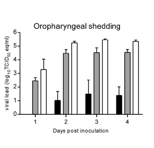
As previously stated, zoonotic diseases are spread from animals to humans via a reservoir host. However there can also be an intermediate host, in which the virus amplifies itself. However this intermediate host is not immune to the disease. Through analysis of urine and saliva collected from a suspected reservoir host from the original outbreak in Malaysia, the reservoir host has been identified as the Pteropus fruit bat . NiV infection has been found in 5 of 14 fruit bat species, with the highest being Pteropus hypomelanus with a 31% infection rate 9. Despite having a relatively high infection rate, the species of Pteropus fruit bats are not susceptible to the virus. The virus is primarily transmitted via body secretions or partially eaten fruit. Pigs have been found to be particularly susceptible to NiV as well as highly contagious to each other. Pigs have been identified as an intermediate and an amplifying host. An amplifying host is defined as a host in which the pathogen can become so prevalent a vector, such as a mosquito, can become infectious. Pigs transmit NiV differently than bats in that they shed the virus primarily by coughing and the expulsion of respiratory secretions and saliva. NiV has not been isolated in pig urine. Pigs are primarily infected directly from Pteropus fruit bats or other pigs. Humans have been found to be infected by three different pathways, Pteropus fruit bat to human, pig to human, and or human to human. Most recent transmission of NiV has been a result of human to human transmission through close contact through respiratory secretions or urine. Although not completely understood, the molecular mechanism of Nipah virus transmission is hypothesized to be by fomites. Fomites are non-living objects which are able to carry infectious material like vectors. Currently this is the main hypothesis because of the increased number of NiV infections being picked up in hospitals. It is unknown how long the virus can survive outside a host. Because of the heavy prevalence of virus transmission via respiratory secretions it is assumed the main site of replication occurs in the tonsils of an infected host 6. This is supported by research done in the transmission of NiV in hamsters. de Wit et al (2011) observed viral shedding of hamsters inoculated with three different doses of varied strength of NiV [FIGURE 3]. The researchers then examined varied forms of shedding of the virus nasally, oropharyngeally, urogenitally, and rectally. The results show, regardless of the dosing, all three dosages resulted in positive viral loads in the oropharyngeal region as soon as two days post inoculation 3 [FIGURE 3]. The fact that the reservoir and intermediate hosts are Pteropus fruit bats and pigs, respectively, is a primary concern for researchers. Pteropus fruit bats are a migratory species capable of flying long distances. Pigs are also a primary export of Southeast Asia as well as a popular farm animal in the same region. The combination of these two key aspects of the Pteropus fruit bat and pigs makes the transmission of the Nipah virus to other parts of the world extremely easy. NiV infections have also been identified in cats, dogs, and horses 9.
FIGURE 3: Data collected in observing the shedding of Nipah virus RNA in inoculated hamsters. Black bars indicate smallest dosage, grey bars was the middle range dosage, white bars was the highest dosage. All differences from days post inoculation were significant. Swabs were taken oropharyngenally. de Wit et al. 2012 http://www.plosntds.org/article/info%3Adoi%2F10.1371%2Fjournal.pntd.0001432
MALAYSIA/SINGAPORE OUTBREAK TRANSMISSION OF NiV
It is believed the transmission from the Pteropus fruit bats to an intermediate host is due to a growing overlap in habitats between the two species [FIGURE 5]. Malaysia is home to a large number of both pig farms and fruit orchards, often located relatively close to each other. The close proximately is thought to have allowed Pteropus fruit bat urine, feces, and or infected fruit to drop onto the pigs or into their habitat. It was speculated pigs had become an amplifying intermediate host because of the enormous death toll in the pig population as well as a majority of the humans infected had a large amount of contact with pigs. In the years after the outbreak NiV antibodies were identified in pig populations in Malaysia. As stated above, a large majority of humans infected had direct contact with pigs. The virus was also able to spread from farm to farm through transportation of unidentified infected pigs. Most likely the workers whom were infected can in direct contact with saliva or other respiratory secretions from an infected pig.13
BANGLADESH/INDIA OUTBREAK TRANSMISSION OF NiV
Because of the variation in the effect and fatality rate of the Nipah virus outbreak in Bangladesh and India [FIGURE 5], a slightly altered transmission path is suggested. It is suggested a more direct path was taken from the reservoir host, Pteropusfruit bats, directly to humans. The large consumption of date palm sap by humans is looked to be where the virus was picked up. Pteropus fruit bats feed heavily on date palm trees, licking the sap which contaminated the sap. Improper cooking of the sap resulted in humans ingesting the infected sap. A second mode of transmission is through contact with infected Ptreopusfruit bat feces, urine, or saliva. Although human to human transmission had not been suggested in the Malaysia/Singapore outbreak, it is believed to have been extremely prevalent. It is believed nosocomial transmission, passing of a pathogen in a hospital setting due to improper hygienic/sterilization methods, was the causation in up to 75% of cases 10. Data collected from various hospitals in Bangladesh and India where patients infected with NiV were treated showed a high amount of NiV RNA on hospital surfaces.
REPRODUCTION OF PARAMYXOVIRUSES
As previously discussed, it is hypothesized there are many models of transmission of Nipah virus from species to species. It can also be assumed, because NiV is a paramyxovirus it acts very similar to other viruses within the family, the reproduction mechanism is very similar to influenza. The virion binds and fuses to the surface of a host cell via the F and G proteins. The lipid bi-layers are then melted and the viral nucleocapsid is released into the host cell. The negative sense viral RNA is transcribed to mRNA which acts as a template for more negative sense viral RNA. The viral RNA is used to make the necessary proteins (N,P,M,F,G,L,C,V,W) which congregate near the cell membrane. Once all the necessary proteins are assembled a new viral cell will bud off and infect other host cells [FIGURE 6]. The new viral cells are able to fuse together and create a huge multinucleated cell called syncytia [FIGURE 6]. A major different between the reproduction of paramyxoviruses and influenza is paramyxovirues are strictly reproduced in the cytoplasm.
PATHOLOGY
FIGURE 6: Cultured Nipah virus. The small arrows indicate the nucleocapsids and the large arrow heads indicate budding virions developing from the cell. Picture scale is 2um. This picture was taken courtesy of Dr. K.B Chua for the use of W.K. Thong in his thesis for the University of Malaya. (Thong 2008)http://dspace.fsktm.um.edu.my/bitstream/1812/591/1/Thesis%20Full%20090711.pdf
Broadly, the Nipah virus causes a multitude of problems within the infected host. However NiV presents itself in a varied set of symptoms depending on the infected species. In general the main mode of dissemination with each host, regardless of species, is by inducing syncytial cell formation [FIGURE 6]. These large multinucleated cells then spread rapidly through the vascular tissue of the infected host. Incubation time in pigs varies but is usually short between 2 and 10 days. The Nipah virus primarily attacks the respiratory system, which is supported by the finding of high concentrations of viral antigens are found in the respiratory tract and lung epithelium 2. Pigs infected with NiV have shown to have an acute fever with symptoms of a respiratory infection which produces a “barking cough”. This “barking cough” has been identified as a telltale sign of Niv infection in pigs. Niv infection also produces signs of nervousness, twitching and trembling, hemorrhaging, and lesions in both the brain and lungs. Although very virulent and contagious among pigs, the mortality rate is very low at roughly 1-5% 2. Unlike Nipah virus infection in pigs, human infection is particularly encephalitic. The onset of a fever after and incubation period as short as two days or as long as a month is the first sign of infection. Common signs of a viral infection such as headache and drowsiness occurred along with the fever. Doctors are able to distinguish a Nipah viral infection by the distinctive symptoms of encephalitis, vasculities, neurological deficits due to necrosis, thrombosis, and ischemia 13. The cerebrospinal fluid is also heavily impacted with an increase of proteins and dead cells present. The heavy encephalitic nature of NiV infection in humans also results in brain lesions, often times causing a relapse of encephalitis in patients who had recovered from NiV infection. Humans infected with NiV have a much higher fatality rate ranging from 40% to 75% depending on the location of the infection 12.
DETECTION AND TREATMENT OF NiV
There has yet to be a standard protocol in detecting Nipah viral infections but the most common process used currently is virus isolation from tissue samples. In all species NiV can be detected and isolated from the kidneys, cerebrospinal fluid, and the liver. Polymerase chain reaction, enzyme-linked immunosorbent, and immunofluorescence assays are also viable detection strategies 14. Currently there are no vaccines or drugs which can cure or treat a NiV infection. The primary approach is to treat the symptoms as best as possible in hope to control the infection. Creating a vaccine is currently extremely difficult because the mutation rate of RNA viruses is extremely high as is most zoonotic diseases, of which Nipah virus is both 9. However several studies have been conducted in targeting the F and G proteins which would inhibit binding of the NiV cells to any host cell 9.
CONCLUSION: THE FUTURE OF NiV
Several steps have been taken in the right direction towards eradicating this extremely harmful disease. After the initial outbreak of Nipah virus in 1998, the Malaysian and Singaporean governments developed a two phase plan in hope to control any future outbreak. Phase one was set to eliminate a majority of the pigs present within the country. Phase two introduced an antibody testing protocol to regulate and observe farms which may be of high risk for an outbreak 7. Both countries also banned the transportation of pigs within the respective countries as well as initiated an educational program to aid the farms in proper handling and the virus itself. As previously stated some initial ground work has also been laid in identifying a possible vaccine which inhibits the activity of F and G proteins on the viral cell 9. Despite these positive steps, Nipah virus should be at the top of the list of major concerns for the human race for several reasons:
1.) Increased human to human transmission in recent outbreaks: Transmission of NiV has now become a concern in many hospitals in Southeast Asia. There have been several cases of doctors becoming infected from treating patients as well as infections resulting from contact with corpses. The more recent outbreaks in Bangladesh and India are suggested to have an estimated 75% or more of the known infections resulting from humans to humans transmission. The main mode of transmission from human to human is hypothesized to be through respiratory secretions and close contact. This mutated strain of NiV has the potential to be extremely detrimental in densely populated cities.
2.) Rising fatality rate in humans: The most recent outbreak in Bangladesh in February of 2013 resulted in increasing the overall fatality rate of Nipah virus infection in Bangladesh to 77% 4.
3.) Lack of knowledge of molecular mechanisms of infection: The molecular mechanisms of how the virus is passed from species to species are still fairly unknown.
4.) Shared habitats: With a rapidly growing human population in the world, particularly in Southeast Asia, there is an increase of overlapping of habitats between humans, pteropus fruit bats, and pigs. This only increases the chance of transmission of NiV between the species as well as the risk of more outbreaks.
5.) Pteropus fruit bat migration and pig dependence: Pteropus fruit bats are migratory animals which can survive in a wide range of environment, while much of rural Southeast Asia is dependent on their pig farms as a source of income and food. The combination of these two aspects opens up many pathways to many new countries and new populations of humans with no previous exposure to NiV.
6.) NiV is an RNA virus and a zoonotic virus: RNA viruses have a high mutation rate which enables them to keep a leg up on both vaccines and host immune systems. Zoonotic viruses also have a high mutation rate. Because NiV is both of these, it is hypothesized it has an extremely high rate of mutation.
eSight is an amazing technological breakthrough – electronic glasses that let the legally blind actually see.
It is the only clinically validated device, in existence, that enables those living with vision loss to see, be mobile, and engage in virtually any Activity of Daily Living.
This device is worn like a normal pair of glasses, and, remarkably, restores sight for someone who is visually impaired.
Most importantly, eSight requires no surgery. Almost instantly after putting them on, an individual with legal blindness or low vision can see in virtually the same manner as someone who is fully sighted. eSight is registered with the FDA and EUDAMED, and is inspected by Health Canada. It is also the only clinically validated wearable technology of its kind.
Neuroscientist Sheila Nirenberg received a MacArthur Genius Award for figuring out, for the first time ever, how our retinas take images from the outside world and turn them into a neural “code” that the brain can understand. It started as a pure research project, but now she’s building the code into a device that could bring sight to the blind.
How does this revolutionary technology actually work?
After putting the electronic glasses on, eSight allows the wearer to see, almost instantly and in beautiful clarity.
In the most simplistic sense, eSight works in three steps. The high speed, high-resolution camera in the center of the device captures what a user is looking at in real time. This video feed is sent into a powerful computer in the housing of the glasses and is enhanced using proprietary algorithms. The feed is then projected in colour on the two near-to-eye OLED screens with unprecedented clarity and virtually no latency or delay.
eSighters can then optimize what they are looking at by using the remote to adjust the color, contrast, focus, brightness and magnification (24x) features. Not only does eSight let wearers actually see, but it also allows them to be truly mobile using the patented Bioptic Tilt Capability. eSighters can tilt the eyewear device to the ideal position for them that can allow the best view of the video feed while maximizing their natural peripheral vision. This, along with short latency, ensures that the eSighter’s balance is not disturbed and no nausea occurs – a common problem faced with immersive technologies such as virtual reality headsets.
Another fun feature about eSight, is it allows individuals to take pictures, and stream video and games by plugging into a laptop, TV or tablet with an HDMI cable, or connecting with Bluetooth or WiFi. That way, whether it is streaming a favourite series at home, taking pictures of notes on the classroom board, or whipping through emails at the office, our eSighters can always be connected.
Who does eSight work for?
eSight works for the overwhelming majority of individuals with vision loss.
Today, our current eSighters live with a variety of conditions, including (but not limited to): Aniridia, Cataracts, Coloboma, Cone-Rod Dystrophy, Diabetic Retinopathy, Glaucoma, Ocular Albinism, Macular Degeneration, Retinopathy of Prematurity (ROP), Stargardt’s Disease, Optic Neuritis, Retinal Detachment, and many more.
According to the World Health Organization, there are approximately 253 million people in the world who are blind. Less than 15% of this population are profoundly or totally blind. Unfortunately, eSight cannot currently help these individuals. However, eSight can work for most of the remaining 85% of this population.
All of our eSighters come from a variety of walks of life. Our youngest user is four years old, and our oldest user is 101 years old. We are also proud to say that our eSighters are spread across over 42 different countries around the world, and counting.
Although all of our eSighters are different, they are united by their fundamental right to see; and eSight is dedicated to making that possible.
What can I do with eSight?
The short answer: virtually anything.
Not only does eSight enable people with vision loss to actually see, but it also restores their independence, confidence, self-esteem and freedom. With eSight, individuals can do almost anything they have only ever dreamt of with their newly restored sight.
With eSight, individuals can participate in virtually all Activities of Daily Living (ADLs). Here are a couple of things that some of our eSighters have been up to:
Seeing the faces of loved ones, in some cases for the first time
Excelling in school and university from being able to see the board from anywhere in the classroom
Plugging into their laptop to work directly from their eSight screens
Catching up on their favourite TV shows
Reading endless amounts of books
Traveling alone to some of the places on their bucket list
Being able to go back to work to help support their family
Watching their favorite sport teams
Pursuing their love of painting, drawing and sketching
Cooking meals for themselves and their loved ones
Going for a walk by themselves
Playing sports with their friends and family
Picking up previously abandoned hobbies (cards, woodworking, etc.)
If you’re going through a stressful period in your life, it can have a huge impact on both your mental and physical health. Being stressed can lower your immunity, making you more susceptible to bacteria and viruses.
What’s chronic stress?
If you’ve been stressed for a long period of time, you have what’s known as chronic stress. As well as affecting your immune system it can lead to depression, anxiety, high blood pressure, sleep problems and increase your chances of suffering from a heart attack or stroke.
Stress and your immune system
Having chronic stress can result in you developing inflammatory and autoimmune disorders. When stressed, your body produces more of the hormone cortisol which can cause your body to struggle to regulate its inflammatory response and attack itself.
Your immune system will be further impaired by your body not producing enough lymphocytes (white blood cells). They are a vital part of your immune system as they fight off bacteria and viruses. Digestion is also impaired whilst you’re stressed and this can lead to gastric ulcers.
How to reduce your stress levels
There are lots of reasons why you might be feeling stressed. It could be that you’ve got a stressful job, you’re the main carer for a sick relative, you’ve got money problems or you’ve been simply doing too much. It’s important to recognise what’s causing you stress and try to tackle it if you can. You might benefit from taking a relaxation course, getting advice from a professional or finding a friend you can confide in. For the sake of your health, you need to take action.
What the Research Shows
Stressed out? Lonely or depressed? Don’t be surprised if you come down with something. Psychologists in the field of “psychoneuroimmunology” have shown that state of mind affects one’s state of health.
In the early 1980s, psychologist Janice Kiecolt-Glaser, PhD, and immunologist Ronald Glaser, PhD, of the Ohio State University College of Medicine, were intrigued by animal studies that linked stress and infection. From 1982 through 1992, these pioneer researchers studied medical students. Among other things, they found that the students’ immunity went down every year under the simple stress of the three-day exam period. Test takers had fewer natural killer cells, which fight tumors and viral infections. They almost stopped producing immunity-boosting gamma interferon and infection-fighting T-cells responded only weakly to test-tube stimulation.
Those findings opened the floodgates of research. By 2004, Suzanne Segerstrom, PhD, of the University of Kentucky, and Gregory Miller, PhD, of the University of British Columbia, had nearly 300 studies on stress and health to review. Their meta-analysis discerned intriguing patterns. Lab studies that stressed people for a few minutes found a burst of one type of “first responder” activity mixed with other signs of weakening. For stress of any significant duration – from a few days to a few months or years, as happens in real life – all aspects of immunity went downhill. Thus long-term or chronic stress, through too much wear and tear, can ravage the immune system.
The meta-analysis also revealed that people who are older or already sick are more prone to stress-related immune changes. For example, a 2002 study by Lyanne McGuire, PhD, of John Hopkins School of Medicine with Kiecolt-Glaser and Glaser reported that even chronic, sub-clinical mild depression may suppress an older person’s immune system. Participants in the study were in their early 70s and caring for someone with Alzheimer’s disease. Those with chronic mild depression had weaker lymphocyte-T cell responses to two mitogens, which model how the body responds to viruses and bacteria. The immune response was down even 18 months later, and immunity declined with age. In line with the 2004 meta-analysis, it appeared that the key immune factor was duration, not severity, of depression. And in the case of the older caregivers, their depression and age meant a double-whammy for immunity.
The researchers noted that lack of social support has been reported in the research as a risk factor for depression, an insight amplified in a 2005 study of college students. Health psychologists Sarah Pressman, PhD, Sheldon Cohen, PhD, and fellow researchers at Carnegie Mellon University’s Laboratory for the Study of Stress, Immunity and Disease, found that social isolation and feelings of loneliness each independently weakened first-year students’ immunity.
In the study, students got flu shots at the university health center, described their social networks, and kept track of their day-to-day feelings using a handheld computer (a new technique called “momentary ecological awareness”). They also provided saliva samples for measuring levels of the stress hormone cortisol. Small networks and loneliness each independently weakened immunity to a core vaccine component. Immune response was most weakened by the combination of loneliness and small social networks, an obvious health stress facing shy new students who have yet to build their friendship circles.
What the Research Means
Emerging evidence is tracing the pathways of the mind-body interaction. For example, as seen with the college students, chronic feelings of loneliness can help to predict health status — perhaps because lonely people have more psychological stress or experience it more intensely and that stress in turn tamps down immunity. It’s also no surprise that depression hurts immunity; it’s also linked to other physical problems such as heart disease. At the same time, depression may both reflect a lack of social support and/or cause someone to withdraw from social ties. Both can be stressful and hurt the body’s ability to fight infection.
All of these findings extend what we know about how stress management and interpersonal relationships can benefit day-to-day health, doing everything from helping us combat the common cold to speeding healing after surgery. The research is in synch with anecdotal reports of how people get sick in stressful times, but understanding exactly howpsychology affects biology helps scientists to recommend the best ways we can build up immunity.
How We Use the Research
Managing stress, especially chronic or long-term stress (even if it’s not intense), may help people to fight germs. When burdened with long-term stressors, such as caring for an elderly parent or spouse with dementia, health can benefit from conscientious stress management.
Kiecolt-Glaser and Glaser confirmed this hopeful option by comparing the immune function of exam-stressed medical students given hypnosis and relaxation training with that of students without training. At first, the immune responses of the two groups appeared to both go down. However, closer inspection revealed that some students took this exercise more seriously than others. Those who didn’t take relaxation training seriously didn’t fare so well; those who practiced conscientiously did actually have significantly better immune function during exams than students who practiced erratically or not at all.
Finally, the newest findings on social stress underscore the value of good friends; even just a few close friends can help someone feel connected and stay strong. Social ties may indirectly strengthen immunity because friends – at least health-minded friends — can encourage good health behaviors such as eating, sleeping and exercising well. Good friends also help to buffer the stress of negative events.
Researchers think the molecule might be better at keeping up with evolving bacteria.
MRSA Researchers at IBM are working on a synthetic molecule to fight antibiotic-resistant bacteria. One of which is MRSA, pictured above. -CDC
When Alexander Fleming discovered penicillin in 1928, the finding was key for two reasons: First, obviously, doctors finally had a way to treat illnesses like pneumonia, gonorrhea, and rheumatic fever. Until then, the approach was to watch, wait, and hope the patient’s immune system cleared the infection; that often didn’t work out. And second, the discovery introduced the idea that we could use molecules found in bacteria and fungi to kill other bacteria—ones that cause infection and illness.
Since then, researchers have been on the hunt to find novel molecules, similar to penicillin, to treat the various bacteria and fungi that infect us. And, from the beginning, it’s been a race against time. Bacteria evolve quickly, and while our goal is to annihilate all of them, their goal is precisely the opposite: To survive at all costs. Research shows that in this tug-of-war effort, humans are being gradually draggedcloser and closer to a bacterial victory. In May 2016 the Review on Antimicrobial Resistance, a research group funded by the UK Department of Health, estimated that 700,000 people die each year from antibiotic resistant infections (these are bacteria that no currently available antibiotics are able to kill). By 2050, an estimated 10 million people could die from this resistance if researchers don’t find a way to keep up with ever-evolving bacteria.
Scientists are employing countless approaches to avoid this outcome. And while most involve finding new molecules or protein in bacteria or fungi, similar to the way Fleming found penicillin, researchers at IBM are taking a different approach: They’ve created a synthetic molecule that works in a novel way to kill each bacterium from the inside out.
The researchers set out to address the scariest of antibiotic resistance scenarios: When a resistant strain of bacteria becomes systemic, spreading through the blood to every organ system in the body. They designed molecules to fight against five of the most drug-resistant strains commonly acquired in hospitals, which often become systemic and lead to organ failure.
Researchers have been working on creating synthetic molecules for some time now, but it’s been difficult. The synthetic molecule needs to be able to biodegrade—it can’t remain inside the body forever—and it also needs to effectively fight bacteria in a way that doesn’t negatively affect other organ systems in the body. Existing drugs that kill highly resistant bacteria typically do so in exchange for toxicity to the liver and other organs.
“We are trying to emulate the exact way that our innate immune system works,” says James Hedrick, a researcher at IBM. He and his team published their findings in a paper out this week in the journal Nature Communications. Our immune systems target a microbe and lyse its membrane, he says—we destroy cellular invaders by breaking down their protective barriers. “When you get an infection, right away your body secretes antimicrobial peptides, which is simply a fancy word for a polymer.” (A polymer, by the way, is also just a fancy word for a big molecule.) In recent years, many scientists have focused on creating these big molecules in the lab.
The problem with using that exact method when you have a systemic infection, Hedrick says, is that when you explode a bacterial cell in the body, it releases its toxins into the bloodstream. That wouldn’t be a terrible thing in isolation. But when you have millions of these dangerous little guys, toxins start to add up.
In the past, Hedrick says, synthetic polymers employed a similar method, where they would essentially explode each bacterium; obliterate it. But instead of causing the bacteria to explode, the new synthetic polymers kill each bacterium from the inside out.
On top of that, Hedrick and his team also think these types of antibiotics will lead to less antibiotic resistance. The polymer works through electrostatic interactions—a positive and negative charge are attracted to one another. But it attracts itself to multiple locations on the bacteria’s surface. This means that even if the bacteria evolves, it’s still highly likely that the bacteria-fighting polymers will remain attracted to one area of the bacteria.
The IBM team has found that the polymer is completely biodegradable and works extremely fast. “What makes this new class of materials so beautiful is that after three days, it degrades completely. It basically just comes in, kills the bacteria, degrades, and leaves.”
So far, all of their studies have been done in mice, but Hendrick says his team is ready to move to human clinical trials. For IBM, that means partnering with a pharmaceutical company to bring the polymer into clinical trials and, potentially, develop it into a drug.
This is all very promising. But the research still has a long way to go before it reaches a doctor’s prescription pad. Even though they’ve showed good results in mice, those same positive effects may not translate to humans, at least not with such efficacy. Most crucial is that the molecules biodegrade as well in humans as they do in mice. Concerns over the long term build-up of such antibiotic polymers in the body has hindered previous development attempts.
There’s also the concern of cost. A lab-engineered polymer will likely be far more expensive to manufacture than traditional antibiotics, and thus potentially more costly for pharmaceutical companies and consumers.
Even if the new treatment proves successful, this is by no means a reason to give up on other efforts to identify post-antibiotic options—as well as those to slow the progress of antibiotic resistance. Things like reducing the number of unnecessary cesarean sections, avoiding using antibiotics for infections that don’t respond to them (like the common cold or the flu) or that a person’s immune system will likely clear without help, and cutting down on their use in meat production will all help to stave off the increasing number of antibiotic resistant bacteria infecting us.
And while the drug may be able to stave off bacterial resistance for some time (Hendrick says it’s hard to predict for how long, exactly), that doesn’t mean it will work forever. “These are really clever bacteria. I am certain that over decades, they will figure out a way to elude the therapy,” says Hedrick. “That’s why this is a never ending kind of fight.”
The Indian Institute of Science in Bengaluru has come up with many useful innovations in science, technology and healthcare. These have made detecting heart conditions and malaria, purifying water, and fixing cataract not just easy, but also affordable.
In the last decade, Indian Institute of Science, in Bengaluru, has churned out a lot of innovations, with the focus being on producing indigenous research and making them available to the country and the world at an affordable rate. From detecting heart conditions to making lenses affordable for cataracts patients, innovations in water purifying and discoveries in cancer drug treatments, here are eight additions to the future of science and technology in India:
Water Purification at a Nanoscale Level
In 2015, Dr. Suryasarathi Bose, Assistant Professor of Department of Materials Engineering and a team invented a water purifying system that could even eliminate harmful bacteria at a nanoscale level. The filter consisted of a porous membrane made of two polymers, along with minute quantities of silver, titanium dioxide and carbon nanotubes. The pores filter out the micron-sized bacteria, while the silver-titanium-carbon mixture kills the bacteria.
A Solar Water Purifier
Another twist to the water purifier, this innovation by Professor Vasant Natarajan, from the Department of Physics is low cost and does not require membranes or electricity. According to Natarajan, this device could purify all kinds of water – sea, bore well, ponds, even rain water – into drinkable water, and produce 1.5 litres out of 3 litres of impure water. Explaining how the device works, he said that first the water is evaporated using solar energy, and then the vapours are condensed on a cold surface. What’s left behind is all the impure substances such as bacteria, arsenic, and fluoride.
A Non-hazardous Stain for Scientists
Researchers in labs often work with a number of chemicals and hazardous materials that could affect their health. Acid stains are used to test a number of chemicals that is probably carcogenic. In March 2016, J Fathima Benazirdeveloped a stain that, if replaced with acid stains, could help researchers reduce their exposure to harmful chemicals. The new stain called Tinto Rang is made from plants, and is even safe for consumption. This indigenous invention could also be the safest in the world, according to Benazir.
Non-invasive Heart Condition Detector
A non-invasive device that can measure heart and lung, called the Fibre Bragg Grating Heart Beat Device, was invented by S Asokan, Professor at Department of Instrumentation and Applied Physics and his team. The device simply needs to be wrapped around a person’s chest, while the sensors detect cardiac activities, measure blood pressure, count blood glucose levels, and monitor respiration. Made of an optical fibre sensor, this device can easily help detect heart conditions early.
A Vaccine to Combat Hepatitis C
In India, 20% chronic liver disease has one cause: hepatitis C virus, which spreads through blood contact, and affects 12 million people. It causes severe liver problems, sometimes even ending up in cancer. In February this year, a team of scientists led by Professor Saumitra Das developed a vaccine that could produce the antibodies to fight the virus. Right now, the vaccine is still being tested on animals, but the results are promising, according to Das.
Smartphone-Turned-Malaria-Detector
Ever thought one could detect malaria through a smart phone? Dr Sai Siva Gorthi, from the department of Instrumental and Applied Physics and her team did so. They converted a smartphone into a powerful microscopic device that eliminates the various stages of blood testing to detect malaria. The team replaced the phone camera with high resolution optics of a microscope. The smartphone also has software that studies the images captured through the microscope and tells even a layman whether it has the malaria virus or not. It requires a tiny amount of blood as a sample.
A Revolutionary Cancer Molecule Inhibitor
In 2012, Sathees C Raghavan, associate professor with IISc’s biochemistry department and his team developed a molecule inhibitor, SCR7, which could revolutionise cancer treatment. In 2014, scientists at MIT tested the molecule and discovered its efficiency and potential in becoming an integral part of anti-cancer drugs. The molecule inhibitor binds with the cancer cells to block its DNA from repair, thereby killing the cancer cells. While the drugs are still under research, the fact remains that an Indian team was vital in creating an anti-cancer drug.
Affordable Lens to Give Vision to Cataract Patients
In a life-saving innovation by Professor G. Mohan Rao at the Department of Instrumentation in 2015, many people who suffer from cataract are now able to see. The team developed economical intraocular lenses (IOLs) in their labs that could be affordable for even poor patients. They succeeded, after months of trials, in creating a thin film of ‘tetraflouroethane’ coating on IOL. This IOL replaces the natural lens in the eyes of a cataract patient. So far, IOLs developed abroad were expensive and inaccessible to most Indians. This, however, changed when Rao and his team succeeded in their tests and transferred the technology to AUROLAB, which now produces these lenses.
Stem cells can be coaxed to self-assemble into structures resembling human embryos.
Two years ago, Shao, a mechanical engineer with a flair for biology, was working with embryonic stem cells, the kind derived from human embryos able to form any cell type. As he experimented with ways of getting cells to form more organized three-dimensional structures by growing them in scaffolds of soft gel, he was looking for signs of primitive neural tissue.
What drew his attention was that the cells seemed to change much faster than expected—they arranged themselves rapidly over a few days into a lopsided circle.
What was it? Shao startled Googling to see if he could identify the structure. That’s when he landed on a website called The Virtual Human Embryo and found some microscope photos of ten-day old human embryos shortly after implantation, fused to the uterine wall. There was the beginning of the amniotic sac and, inside it, the embryonic disc, or future body. They matched what he was seeing.
In this microscope movie, filmed over four days, stem cells self-organize in ways that mimic a human embryo.
COURTESY OF UNIVERSITY OF MICHIGAN
Shao informed his coworkers, a mixed team of biologists and engineers, at the University of Michigan. “When I showed the image to the team, everyone said, “Wow, we need to figure out what to do,” says Shao. Had they somehow made a real human embryo from stem cells? “At that point, we started to be more cautious.”
The embryo-like structures, the team soon determined, are not complete and couldn’t become a person. They lack the cell types needed to make a placenta, a heart, or a brain. Even so, the Michigan “embryoids” are realistic enough that the lab has been destroying them using a bath of detergent or formaldehyde to make sure they don’t develop any further.
The work in Michigan is part of a larger boom in organoid research—scientists are using stem cells to create clumps of cells that increasingly resemble bits of brain, lungs, or intestine (see “10 Breakthrough Technologies: Brain Organoids”). Now some like Shao are finding it’s possible to mimic the embryo itself. This year, for example, researchers in Cambridge, U.K., built a convincing replica of a six-day-old mouse embryo by combining two types of stem cells. That group is now trying to do the same with human cells, as are a few others, including one at Rockefeller University in New York. What’s emerging, say scientists, is a new technology, which they call “synthetic embryology,” and which they believe may let them probe the fascinating opening chapters of human development in detail for the first time.
That’s been difficult to do because normal embryos don’t keep growing more than about a week in a lab. Key events after that are largely inaccessible to science: they occur in the darkness of the human uterus even before most women know they’re pregnant.
A microfluidic device used at the University of Michigan to cultivate organoids made from embryonic cells. About 10 organoids can fit inside each of the small blue channels.
COURTESY OF UNIVERSITY OF MICHIGAN
What’s more, research on real human embryos is dogged by abortion politics, restricted by funding laws, and limited to supplies from IVF clinics. Now, by growing embryoids instead, scientists see a way around such limits. They are already unleashing the full suite of modern laboratory tools—gene editing, optogenetics, high-speed microscopes—in ways that let them repeat an experiment hundreds of times or, with genetic wizardry, ask a thousand questions at once.
One result already from the Michigan team: dramatic close-up video of stem cells self-organizing into structures that mimic embryos.
“It’s amazing that [stem cells] have this capability,” says Jianping Fu, the University of Michigan professor in whose engineering lab Shao was a student. He says the emergence of something with an embryo’s shape, and some of its features, was “a complete surprise; I still can’t believe it. But it shows these cells remember what they are supposed to do.”
Scientists at Michigan now have plans to manufacture embryoids by the hundreds. These could be used to screen drugs to see which cause birth defects, find others to increase the chance of pregnancy, or to create starting material for lab-generated organs. But ethical and political quarrels may not be far behind. “This is a hot new frontier in both science and bioethics. And it seems likely to remain contested for the coming years,” says Jonathan Kimmelman, a member of the bioethics unit at McGill University, in Montreal, and a leader of an international organization of stem-cell scientists.
What’s really growing in the dish? There no easy answer to that. In fact, no one is even sure what to call these new entities. In March, a team from Harvard University offered the catch-all “synthetic human entities with embryo-like features,” or SHEEFS, in a paper cautioning that “many new varieties” are on the horizon, including realistic mini-brains.
Shao, who is continuing his training at MIT, dug into the ethics question and came to his own conclusions. “Very early on in our research we started to pay attention to why are we doing this? Is it really necessary? We decided yes, we are trying to grow a structure similar to part of the human early embryo that is hard otherwise to study,” says Shao. “But we are not going to generate a complete human embryo. I can’t just consider my feelings. I have to think about society.”
Other scientists, however, are determined to see just how far the science leads, up to and including forging the first complete human embryo from stem cells. That’s the case of Ali Brivanlou, an embryologist who leads a lab at Rockefeller University, in New York City. “My goal is to maximize the modeling, in vitro, of human development,” Brivanlou wrote in an e-mail. “Therefore, we would like to be as accurate as possible and as complete as possible.”
Taking shape
Embryonic stem cells were first isolated from spare, days-old IVF embryos in 1998 by scientists in Wisconsin. Early on, in its first few days, an embryo is little more than a mass of these identical, blank-slate, cells. Their specialty: making any other type of cell in the body. With an eye toward eventual medical treatments, companies have used them to produce neurons and beta cells that respond to insulin. Left alone in a dish, they’ll spontaneously turn into heart muscle and start beating.
Scientists have started seeking ways to coax stem cells to form more complicated, organized tissues, called organoids. These mini-organs aren’t the real thing. Instead, they’re far smaller—the size of sand grains—and often less sophisticated. But they can still have basic aspects of, say, the branching airways and wavy cilia of a lung. Last year, researchers used brain organoids to show how the Zika virus can infect brain cells.
By 2014, such efforts started yielding evidence that stem cells might, if given the right cues, directly reenact early events in an embryo. Brivanlou’s lab had the idea of corralling stem cells within tiny dots on a micro-patterned surface. Containing the cells helped lead to a surprising effect. They developed an organized “primitive streak”—a feature of a two-week-old human embryo when cells lay down the first hint of a body plan, deciding which side is left which is right.
Those embryoids were not natural. They were thin, grown as a flat sheet, and their streaks were circles, not lines as in a true embryo. “But it worked better than we thought,” says Aryeh Warmflash, a Rice University professor who ran the experiment while working at Rockefeller. “What we have increasingly realized is that the cells are programmed to make an embryo. That is what they want to do. If cells are in the right shape, at the right density, and you give them the right signal, the cells just take over from there, they talk to each other.”
At Michigan, Fu says his lab, working with Michigan biologist Deborah Gumicio, hit on its own method for making embryoids almost by accident while studying whether mechanical signals, like growing cells in a gel that is soft or sticky, could enhance their ability to form certain tissues.
One experiment involved encouraging gut cells to form a lumen, or hollow cyst. As a control experiment, they also cultivated embryonic stem cells in the same way. That is when “serendipity hit,” says Fu. The stem cells polarized into spheres that bore similarity to the start of an amniotic cavity. “[After] that is when we saw all the fascinating self-organizing features,” says Fu.
Ethical questions
Further tests demonstrated that the embryoids represented only a part of the embryo. While they had the beginnings of an amniotic sac, they lacked an entire lineage of cells, called trophoblast, whose role is to make the placenta. And inside the clump of cells that constitutes an embryo proper, the researchers detected only one of three key types needed to make a complete body.
When the team published its findings in early August, they went mostly unnoticed. That is perhaps because the scientists carefully picked their words, straining to avoid comparisons to embryos. Shao even took to using the term “asymmetric cyst” to describe the entities that had so surprised the team. “We have to be careful using the term synthetic human embryo, because some people are not happy about it,” says Fu.
An “embryoid” created from stem cells shares key features with a real human embryo, like an amniotic sac, but lacks other elements.
COURTESY OF YUE SHAO, UNIVERSITY OF MICHIGAN
Currently, scientists in the U.S. and U.K. working with natural human embryos observe a limit on their work called the “14-day rule.” No human embryo is studied beyond two weeks, or past when the primitive streak forms, whichever comes first. Before then, no one thinks they have any kind of sentience and are “incapable of feeling pain” according to the 1984 Warnock Report that enshrined the rule.
For decades, that rule has offered a convenient and clear line in the sand. And the same limits are being applied to embryoids, at least for now. Following guidelines promulgated last year by Kimmelman’s international stem-cell society, Fu’s team destroys the cells just five days after they’re made. This prevents the structures from developing what bioethicists term “features of concern”—such as a primitive nervous system.
But scientists are prepared to argue that their structures aren’t real embryos, and that they should be able to stretch the limit. Some experts are calling for an end to the rule altogether, saying it is outdated. John Aach, a scientist at Harvard Medical School, thinks entirely new ethical measuring sticks will be needed to help guide tests of organoids. For instance, could a mini-brain grown in the lab somehow feel suffering? And can our definition of an embryo withstand evidence that labs can make new sorts never before seen? “All great scientific advances have a way of exposing the imprecision of common concepts and forcing people to rethink them,” says Aach.
Even before his paper came out, Shao was buttonholing ethics experts, including Insoo Hyun, a professor at Case Western University, at a conference this year in Boston. Hyun felt the young researcher was on safe ground because his structure didn’t contain every part of an embryo. “I think that they should design experiments to focus on specific questions, and not model everything,” says Hyun. “My proposal is, just don’t make the whole thing. One team can make the engine, another the wheels. The less ambiguous morally the thing is that you are making, the more likely you can do your research unimpeded.”
There’s yet another reason to be cautious. The U.S. currently bars federal funding for any study of embryos, no matter how they are made, under a law called the Dickey-Wicker Amendment.
While today’s embryoids don’t appear to be covered by the legal restriction, they might be if scientists make them realistic enough. In response to written questions, the science policy office of the National Institutes of Health, the $33-billion-a-year funding agency, says it has an internal process it uses to analyze grants and to determine if “proposed research would create an organism that meets the statutory definition of a human embryo.”
The Michigan scientists, whose project used funds from two NIH grants, say agency officials haven’t raised any objections so far. For now, the embryoids live and die in boxes made of lucite and metal and are fed with culture medium. “Because of the really heavy engineering component to these entities, I think you will be able to argue these are not organisms,” says Hyun. That’s a point that Shao has sought to emphasize, too. When Shao presented the group’s work this year, he added to his slides an ethics statement outlined in a bright yellow box, saying the embryoids “do not have human organismal form or potential.”
Scientists are trying to manufacture eggs and sperm in the laboratory. Will it end reproduction as we know it?
But such definitions could be a moving target. The whole point of the structures is the surprising, self-directed, even organismal way they develop. Robert Cork is the head of the Virtual Embryo Project, which maintains the images the Michigan team used to identify their structures. When I asked him about Shao’s paper, Cork told me that the embryoids could go on to make some of the parts they’re currently missing, if the experiments were allowed to progress. “This would suggest that if they can keep the cysts viable for longer they might go ahead and start to develop into something more ‘embryo-like,’” says Cork.
High-throughput
Jianping Fu is a professor of mechanical engineering at the University of Michigan.
COURTESY OF UNIVERSITY OF MICHIGAN
Fu says the next step in his Ann Arbor laboratory is to perfect procedures for making embryoids with specific characteristics, and in larger numbers. Initially, of every 100 “cysts” the Michigan scientists grew, only five ended up with the asymmetric shape reminiscent of the amniotic sac. But the they have already learned how to make that shape emerge every time. The production of embryoids will become “programmable and scalable,” Fu predicts.
Drugs could be tested on the embryoids, for instance to flag any that have toxic effects and cause birth defects. Fu’s hope is that synthetic embryology might eventually help engineers grow complete human organs. “I am not talking about a human body without a brain. But what is a true possibility is you could develop a mini-gut or a mini-liver, since the embryo develops them, too. And if you have the primitive organs, they could grow into a functional one,” Fu predicts. The lab has started growing embryoids on a chip about the size of a credit card. Etched into it are six microchannels, each accommodating 10 of the entities, which are suspended in hydrogels and fed nutrients held in miniature buckets. Fu calls it “high-throughput manufacturing.”
This way, he says, “everything can be triggered and under control.”
Artificial mouse embryo after 48 hours (right) and 96 hours, with embryonic tissue in red and extra-embryonic tissue in blue
Sarah Harrison and Gaelle Recher, Zernicka-Goetz Lab, University of Cambridge
Artificial mouse embryos grown from stem cells in a dish could help unlock secrets of early development and infertility that have until now evaded us.
Magdalena Zernicka-Goetz at the University of Cambridge and her team made the embryos using embryonic stem cells, the type of cells found in embryos that can mature into any type of tissue in the body.
The trick was to grow these alongside trophoblast stem cells, which normally produce the placenta. By growing these two types of cell separately and then combining them in a special gel matrix, the two mixed and started to develop together.
After around four-and-a-half days, the embryos resembled normal mouse embryos that were about to start differentiating into different body tissues and organs.
“They are very similar to natural mouse embryos,” says Zernicka-Goetz. “We put the two types of stem cells together – which has never been done before – to allow them to speak to each other. We saw that the cells could self-organise themselves without our help.”
This is the first time something resembling an embryo has been made from stem cells, without using an egg in some way. Techniques such as cloning, as done for Dolly the sheep and other animals, bypass the need for sperm, but still require an egg cell.
Body plan
The artificial embryos are providing new insights into how embryos organise themselves and grow, says Zernicka-Goetz. The team engineered the artificial embryos so the cell types fluoresced in different colours, to reveal their movements and behaviour as the embryos go through crucial changes.
Mammal embryos were already known to start as a symmetrical ball, then elongate, form a central cavity and start developing a type of cell layer called mesoderm, which ultimately goes on to form bone and muscle.
“We didn’t know before how embryos form this cavity, but we’ve now found the mechanism for it and the sequential steps by which it forms,” says Zernicka-Goetz. “It’s building up the foundations for the whole body plan.”
“The work is a great addition to the stem cell field and could be extended to human stem cell populations,” says Leonard Zon at Boston Children’s Hospital, Massachusetts. “Using the system, the factors that participate in embryo development could be better studied and this could help us understand early events of embryogenesis.”
But Robin Lovell-Badge at the Francis Crick Institute in London says that the embryos lack two other types of cell layer required to develop the bodies’ organs: ectoderm, which forms skin and the central nervous system, and endoderm, which makes our internal organs.
Zernicka-Goetz hopes to see these types of cell layers develop in future experiments by adding stem cells that normally form the yolk sac, a third structure involved in embryonic development, to the mix.
Hidden steps
If a similar feat can be achieved using human stem cells, this could tell us much about the earliest stages of our development. Current research is limited by the number of excess embryos that are donated from IVF procedures. But the new technique could produce a limitless supply, making it easier to conduct in-depth research. These artificial embryos may also be easier to tinker with, to see what effect different factors have in early embryogenesis.
Disrupting development in this way may provide new insights into the causes of abnormal embryo development and miscarriage. “You would be able to understand the principles that govern each stage of development. These are not normally accessible, because they happen inside the mother,” says Zernicka-Goetz.
But it is doubtful that this work could ever lead to fully grown babies in the lab. Lovell-Badge says the artificial embryos are unlikely to develop in vitro much further than shown in the study, as they would soon need the supply of nutrients and oxygen that a placenta normally channels from the mother.
“We’re not planning to make a mouse in the lab using stem cells,” says Zernicka-Goetz. But she is hopeful that adding yolk sac stem cells will allow these artificial embryos to survive long enough to study the beginnings of organs like the heart.